Holon-S1 Was Never Trained on Osteosarcoma, Yet It Read Key Genes from Pathology Slides
Sophont and MedARC release Holon-S1: it predicts spatial gene expression directly from routine H&E slides, reproduces key molecular findings in osteosarcoma cases it was not trained on, and finds in triple-negative breast cancer that EREG spatial distribution correlates with chemotherapy response. Currently for research use only; open to collaboration.
Tanishq Mathew Abraham announced Holon-S1 on X. The launch tweet was included in venture capitalist Chamath Palihapitiya's newsletter, which Tanishq himself did not expect.
Holon-S1 is a model developed by Sophont and MedARC, with a straightforward goal: predict genes and biomarkers from routine H&E-stained pathology slides.
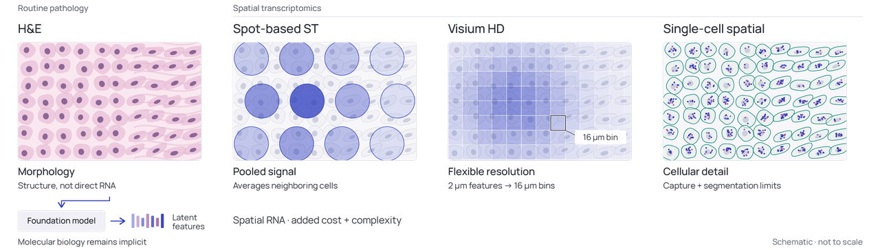
Patients with the same cancer diagnosis often have completely different tumor biology. Molecular testing can reveal these differences, but it is slow, expensive, and hard to scale. Spatial transcriptomics can measure gene expression while preserving location information, but it also has high technical barriers and costs. Holon-S1 aims to provide answers directly from existing slides. It is currently for research use only and has not entered clinical practice.
Video from the Holon-S1 launch tweet.
H&E staining has been the gold standard in pathology since 1877. Digital pathology slides can be converted by foundation models into vector representations (embeddings), but the biology encoded in them is often a black box. Holon-S1 translates these morphological features into spatially resolved gene expression profiles and biological programs. Its outputs can be inspected: which genes, which biological processes, and where in the tumor they are distributed.
Performance: Two 2026 Comparison Models
In the technical report, Holon-S1 was compared with two 2026 models. When predicting the locations of gene activity, its median per-gene correlation was:
- DeepSpot-M: breast cancer 0.49 vs. 0.21; cervical cancer 0.31 vs. 0.11, with cervical cancer not present in the training set
- Path2Space: breast cancer 0.34 vs. 0.17
Path2Space only handles breast cancer, whereas Holon-S1 is a pan-cancer model. It was also evaluated on 16 organs, more than 15 scanners, FFPE (formalin-fixed, paraffin-embedded) and fresh frozen tissue.


Osteosarcoma Case: Reading Key Genes in a Cancer Type It Was Not Trained On
Holon-S1 was never trained on osteosarcoma data. The research team conducted a retrospective N-of-1 analysis using pathology slides publicly shared by GitLab co-founder Sid Sijbrandij. Sijbrandij was diagnosed with osteosarcoma in 2022 and later publicly released a large amount of molecular testing data. His team found a fibroblast program in the tumor microenvironment, with FAP (fibroblast activation protein) as a gene relevant to targeted therapy and high MDM2 expression.
From routine slides, Holon-S1 predicted high expression of fibroblast-related genes in tumor regions, including FAP; predicted MDM2 expression was higher than in 9 of 10 control patients. Post-treatment slides showed increased immune infiltration, consistent with IHC (immunohistochemistry) data.
It should be clear: Holon-S1 is a research tool and played no role in this patient's treatment.

Breast Cancer: EREG Spatial Distribution Correlates with Chemotherapy Response
In a triple-negative breast cancer cohort, Holon-S1 identified a potential new biomarker: the spatial arrangement of EREG gene activity correlates with chemotherapy response. Before this, no one had linked EREG to chemotherapy response in this cancer.
In the IMPRESS discovery cohort, the EREG spatial gradient had an average precision of 0.690, versus only 0.567 for sTIL; in the TransNEO validation cohort, it was 0.754 vs. 0.505. sTIL (stromal tumor-infiltrating lymphocytes) is an immune metric commonly used by pathologists. In the validation cohort, this spatial feature performed more consistently than sTIL.

Interpretability
Many pathology AI models only provide a prediction score without explaining the underlying biology. Holon-S1 maps genes and biological programs onto the tumor, allowing researchers to verify them. It also distinguishes regions dominated by B cells from those dominated by T cells, with a balanced accuracy of 88.5% and an AUROC of 0.943. Conventional morphology has difficulty distinguishing these two types of lymphocytes.
The technical report also includes a cross-scanner stability experiment. For the same prostate slides scanned with Grundium, Hamamatsu, and Leica, the median Wasserstein-1 distance for consistency of Holon-S1's predicted distributions was 0.057, compared with 0.120 for DeepSpot-M. Even when the scanner changed, Holon-S1's predictions were more stable.
Current Status
Holon-S1 is for research use only. The team says prospective validation is still needed before personalized cancer treatment can become more accessible. The full technical report is at sophont.med/blog/holon, authored by Hassan Muhammad, Benjamin Warner, Paul Steven Scotti, and Tanishq Mathew Abraham. The team also welcomes independent evaluation.
Collaboration areas include biopharma, life science teams, and academic research groups. If you have pathology data on hand, you can apply at: sophont.med/holon
Source material comes from Tanishq Mathew Abraham's X post and the Sophont technical report.
发布时间: 2026-10-26 07:38